3D mammography, sometimes called 3D tomography or tomosynthesis, is an advanced mammogram that gives radiologists more information when examining breast tissue. Many women ask about it because it can provide a more complete view than standard mammography alone, especially in areas where breast tissue overlaps.
At Bay Radiology, 3D mammography is part of our advanced Breast Imaging services in Panama City, with every study interpreted by Board Certified, Fellowship Trained radiologists.
What Is 3D Mammography?
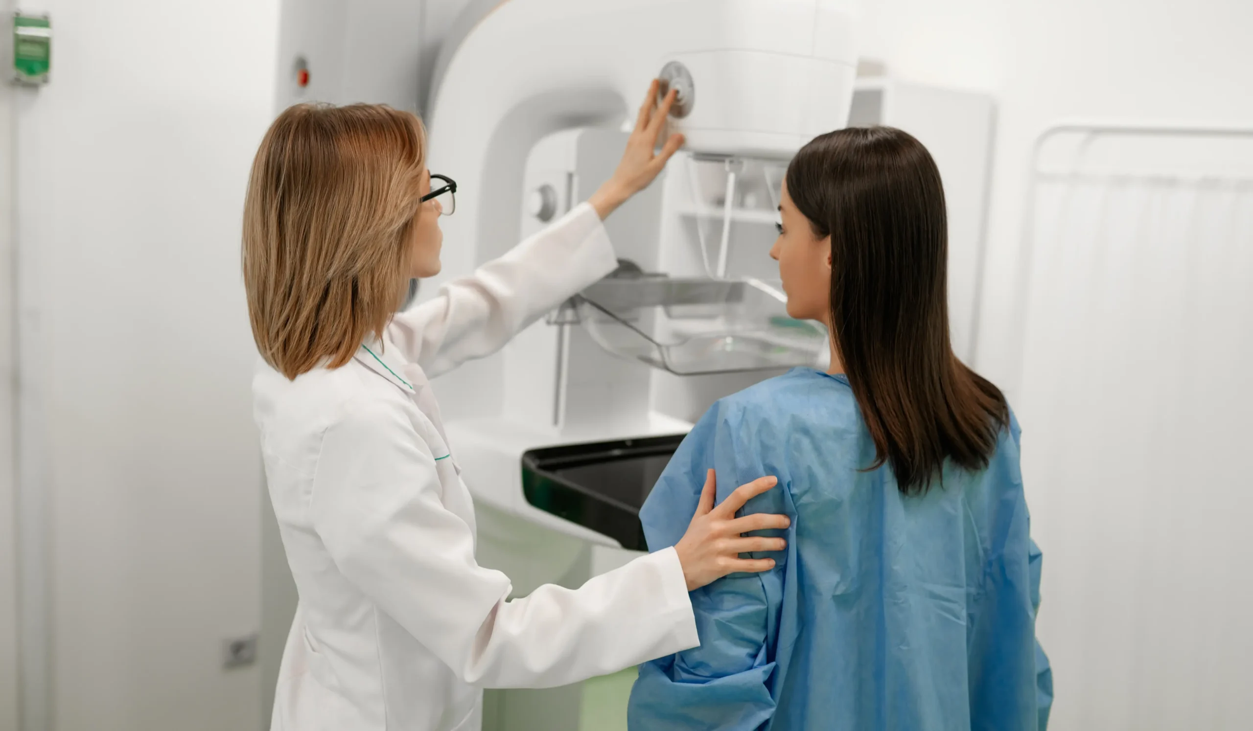
3D mammography is an advanced form of digital mammography that captures multiple low-dose images of the breast from different angles during a single exam. Those images are reconstructed into thin sections of breast tissue that radiologists can review one at a time.
For you as the patient, a 3D mammogram feels very similar to a standard mammogram. The main difference is in how the images are created and how the breast tissue is reviewed afterward.
Benefits of 3D Mammography
3D mammography offers several benefits that can support more informed breast Imaging and reduce uncertainty during screening or follow-up.

Greater Visibility Through Breast Tissue
By showing breast tissue in thin sections, 3D mammography can help separate structures that might otherwise appear stacked on top of one another in a standard image.
Earlier Detection of Subtle Changes
The added image information can help reveal small findings that may be harder to recognize on traditional mammography alone.
Helpful for Women with Dense Breast Tissue
Dense breast tissue can make mammograms more difficult to interpret. 3D mammography gives radiologists additional information when reviewing these studies.
Fewer Follow-up Imaging Exams for Some Patients
In some cases, 3D mammography helps answer questions during the initial exam, which may lower the chance of being called back for more images.
Standard Mammography vs. 3D Mammography
Standard digital mammography is still an excellent screening tool and remains the foundation of breast cancer detection. It has helped women and their providers catch breast changes early for years, and it continues to play a central role in routine breast screening.
3D mammography offers an additional option when more information may be helpful. Rather than replacing standard mammography, it expands what is available so Breast Imaging can be approached with more flexibility based on the patient and the reason for the exam.
At Bay Radiology, both standard digital mammography and 3D mammography are important parts of breast care. The right approach depends on your needs and the recommendation of the provider ordering the study.
Why 3D Mammography May Be Recommended
3D mammography is often used for routine breast cancer screening, not just for diagnostic follow-up. You do not need to have a breast problem or an abnormal mammogram first for this technology to be part of your care. For some patients, it is simply the screening approach their provider recommends based on breast tissue, prior imaging, or personal history.
Routine Screening
3D mammography can be used as part of your regular screening mammogram. For some providers and patients, it is the preferred screening approach.
Dense Breast Tissue
If you have dense breast tissue, 3D mammography may be recommended because dense tissue and some abnormalities can both appear white on a standard mammogram. That can make certain findings harder to separate on a flat image.
Follow-up After a Screening Mammogram
If a screening exam raises a question, 3D mammography may also be used to take a more focused look at a specific area.
Personal or Family History
Your personal health history, family history of breast cancer, and prior breast imaging can all influence the type of mammogram your provider recommends.
If you are due for a mammogram and are not sure what type of screening makes sense for you, it is worth asking about your options. Your provider can help determine whether standard digital mammography or 3D mammography is the better fit for your situation.
What to Expect During a 3D Mammogram
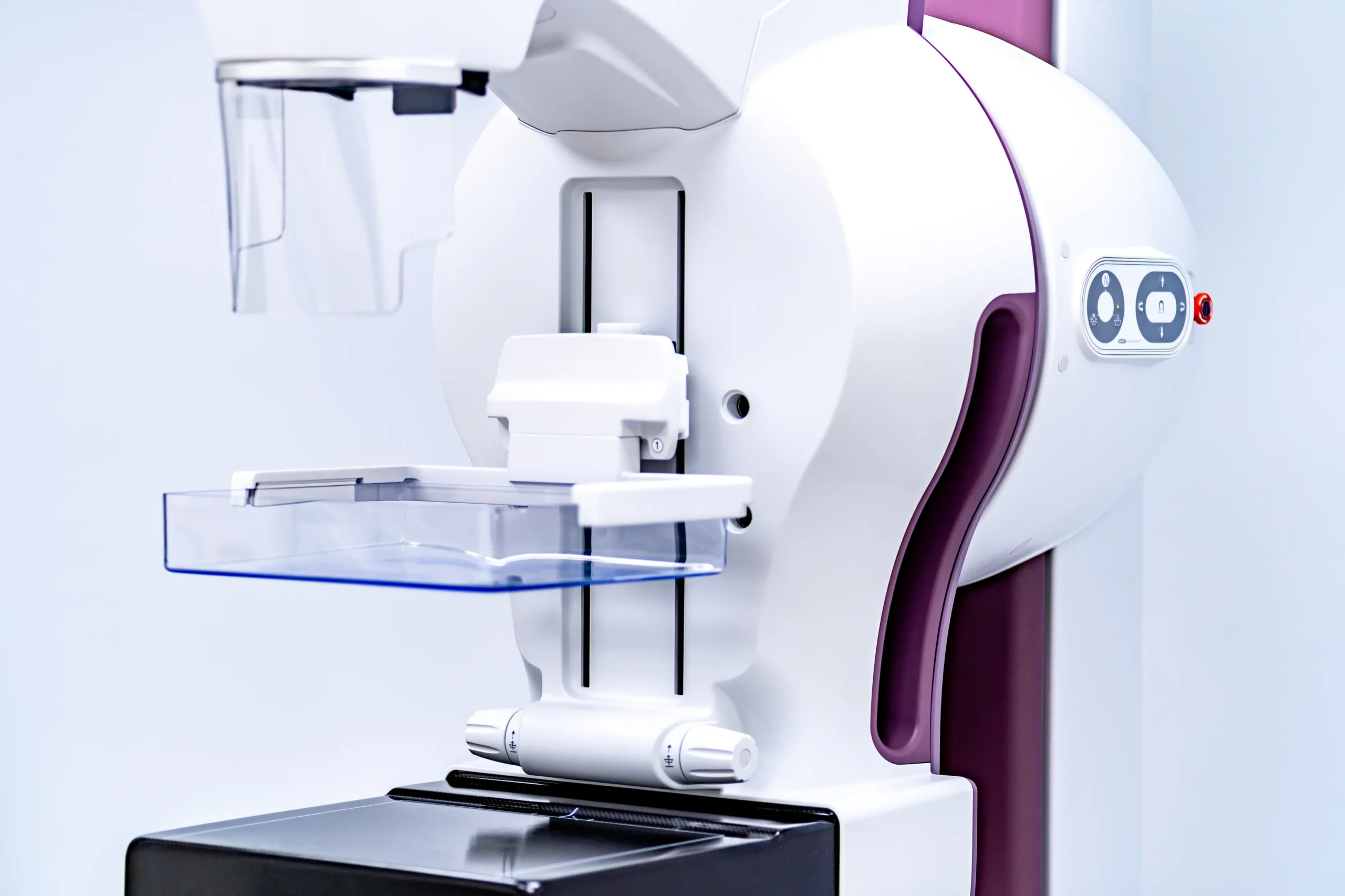
A 3D mammogram feels very similar to a standard mammogram. During the exam, you will stand in front of the imaging system while a member of the Breast Imaging team positions your breast on the imaging plate.
The breast is briefly compressed between two plates so clear images can be captured. This can feel uncomfortable for a few seconds, but it helps ensure the images are accurate and reduces the chance that additional images will be needed.
As the images are taken, the imaging arm moves slightly over the breast to capture a series of pictures. The process only takes a few moments, and most appointments are finished within about 15 to 20 minutes.
After the exam is complete, you can return to normal activities right away.
How to Prepare for Your Appointment
Most 3D mammograms do not require much preparation, but there are a few things worth keeping in mind before your appointment.
- Avoid deodorant, lotion, powder, or perfume on the day of your exam, as these products can appear on mammogram images.
- Wear a two-piece outfit so it is easier to change for the exam.
- Let the Breast Imaging team know if you have any symptoms, such as a lump, breast pain, or nipple discharge.
- Bring information about prior mammograms if they were performed at another facility.
Tell us if you are pregnant or think you might be pregnant before the exam begins.
See More Preparation Tips
Results and Next Steps
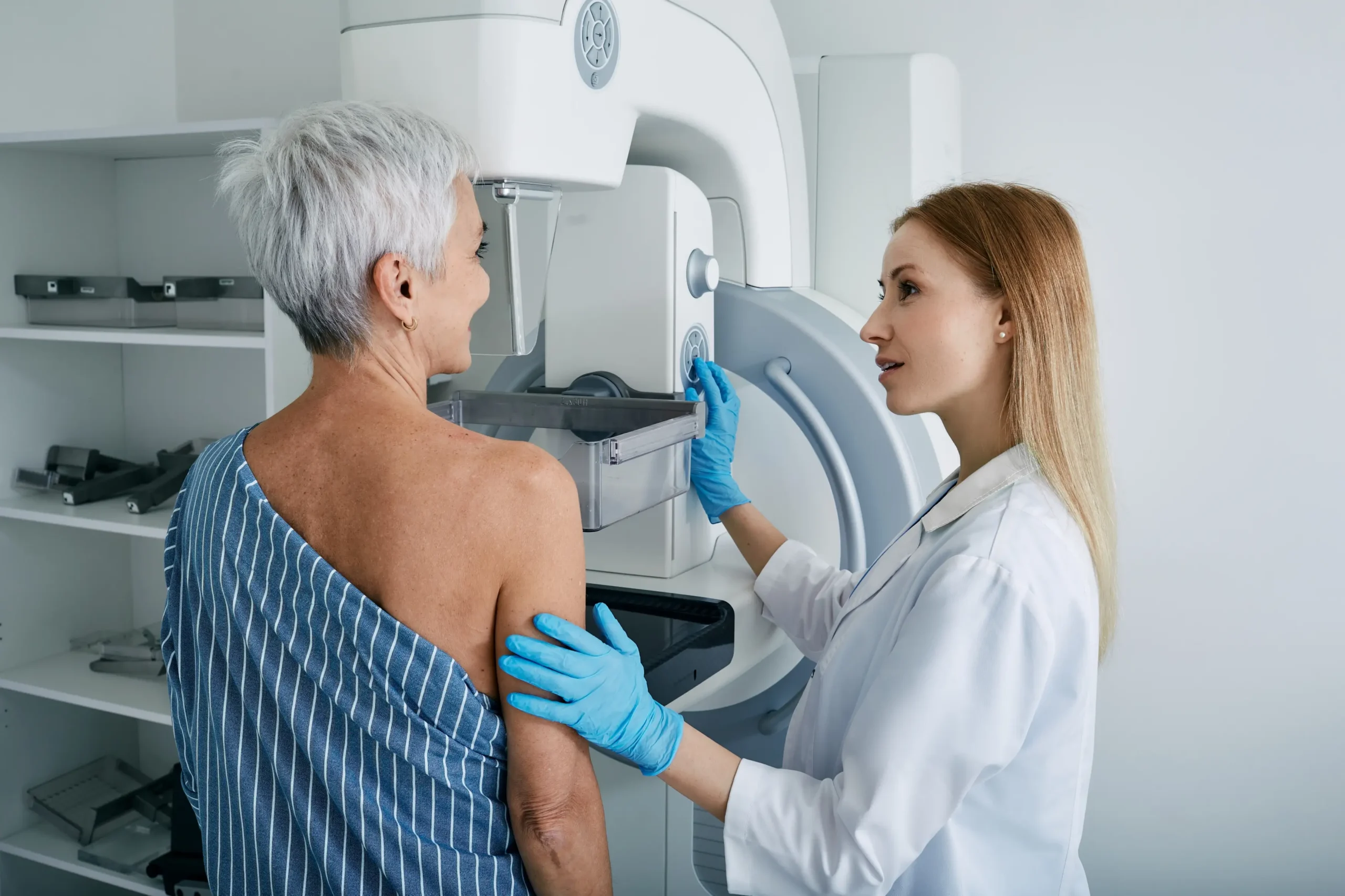
After your exam, your 3D mammogram is reviewed at Bay Radiology by Board Certified, Fellowship Trained radiologists with specialized expertise in breast Imaging.
A report is then sent to the provider who ordered your mammogram. For many patients, the next step is simply returning to routine screening at the interval their provider recommends. If any follow-up is needed, your provider will explain what comes next and why.
Frequently Asked Questions
What is tomosynthesis?
Tomosynthesis is the medical name for the technology used in 3D mammography. You may hear the terms breast tomosynthesis, 3D mammography, or sometimes 3D breast imaging, but they all refer to the same type of exam.
The word tomosynthesis describes how the images are created. Instead of producing one flat image of the breast, the imaging system captures multiple low-dose images from different angles. A computer then reconstructs those images into thin sections of breast tissue that radiologists can review one layer at a time.
This layered view is what gives 3D mammography its name and is why the technology can provide additional imaging detail compared to a traditional 2D mammogram.
Does insurance cover 3D mammograms?
In many cases, yes, 3D mammography is covered by insurance, especially when it is used for routine breast cancer screening. However, coverage can vary depending on your insurance plan and whether the exam is being done for screening or diagnostic reasons.
Because plan details are not always the same, it is a good idea to confirm coverage ahead of time if you are unsure. That way, you know what to expect before your appointment.
How long does a 3D mammogram take?
Most 3D mammogram appointments are fairly quick. The imaging itself only takes a few minutes, and many visits are completed in about 15 to 20 minutes.
Even though the technology is more advanced, the exam does not usually feel much longer than a standard mammogram. Most of the appointment time goes toward positioning and making sure the images are captured correctly.
Does a 3D mammogram hurt?
A 3D mammogram can feel uncomfortable for a few seconds because the breast must be gently compressed to capture clear images. Most patients describe the feeling as pressure rather than pain, and it only lasts briefly while the image is taken.
If you have had a standard mammogram before, the experience is very similar. The Breast Imaging team works carefully to position you as comfortably as possible while still getting the images needed for an accurate exam.
How accurate are 3D mammograms?
Many women ask this question because they want to know whether 3D mammography is actually better or just newer technology. The benefit of 3D mammography is that it gives radiologists more information to work with by showing the breast in thin sections rather than only flat images.
That added detail can help with interpretation, especially in areas where tissue overlaps or where dense tissue makes screening more complex. Like all mammograms, though, it is still one part of your overall breast health evaluation, and results are interpreted in the context of your history, symptoms, and prior imaging.
Is a 3D mammogram better than a regular mammogram?
This is one of the most common questions women ask, and the answer is not simply that one is good and the other is bad. Standard digital mammography is still an excellent screening tool and remains the foundation of breast cancer detection.
3D mammography builds on that by providing additional imaging detail in some situations. For many women, that can be helpful during routine screening, especially when breast tissue is harder to interpret on standard images alone. The right choice depends on your history, your breast tissue, prior imaging, and your provider’s recommendation.
Does a 3D mammogram use more radiation?
This question usually comes up because people hear “more images” and assume that must mean a major increase in radiation. In reality, 3D mammography uses low-dose X-rays, and modern systems are designed to keep radiation exposure within established safety guidelines while still producing high-quality images.
Your provider recommends mammography because the value of screening and early detection far outweighs the low level of radiation used during the exam.
Does a 3D mammogram show lymph nodes?
A 3D mammogram is designed to evaluate breast tissue, not to serve as a dedicated lymph node study. However, depending on your anatomy and how the images are positioned, some nearby lymph nodes, especially in the underarm area, may be visible on the exam.
If lymph nodes need a closer look, that is usually handled with additional breast Imaging such as ultrasound or another recommended study. A mammogram is one part of the larger picture, not the only imaging tool available.
Have Questions Before Your Mammogram?
From preparation to scheduling to understanding the type of exam your provider ordered, Bay Radiology is here to help you feel more informed before your visit.