A breast ultrasound is a common breast Imaging exam used to evaluate a specific area of concern in the breast. It is often ordered when you have a lump, tenderness, or another change that needs to be checked, or when another breast Imaging exam raises a question that ultrasound can help answer.
At Bay Radiology, breast ultrasound is performed as part of our Breast Imaging services in Panama City, with studies interpreted by Board Certified, Fellowship Trained radiologists.

What Is a Breast Ultrasound?
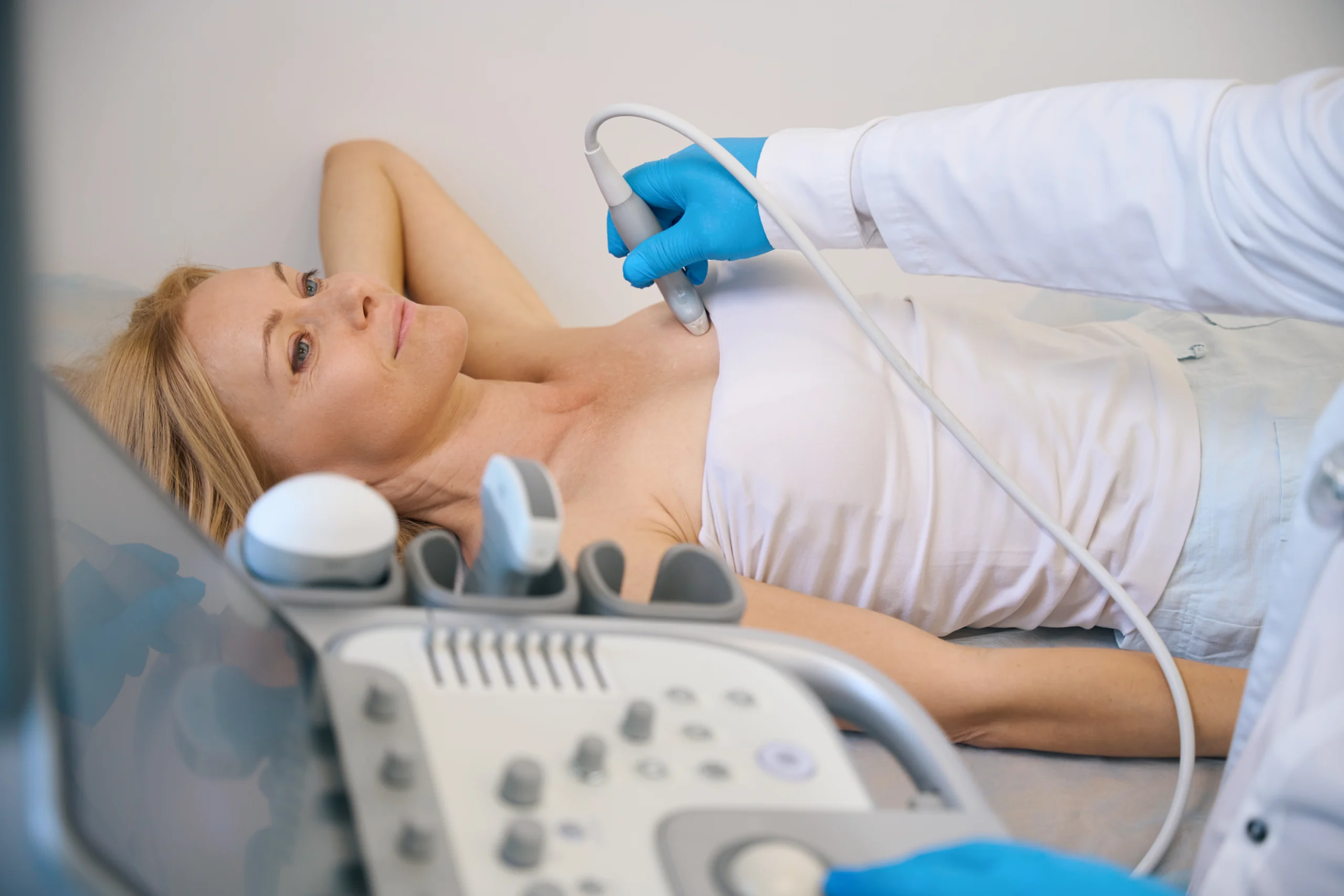
A breast ultrasound uses sound waves to create images of breast tissue. Unlike mammography, it does not use radiation. During the exam, a small handheld device is moved over the skin to capture images of the area being evaluated.
Breast ultrasound is especially useful for looking at breast tissue in real time and helping radiologists understand what a finding may represent, such as whether an area appears solid or fluid-filled.
When Breast Ultrasound Is Used
Breast ultrasound is often used to evaluate breast tissue in ways that are hard to capture on other Imaging exams. It can help:
- Clarify what a finding may be by showing whether an area looks more like a fluid-filled cyst or a solid mass
- Focus on one specific area of the breast when the goal is to evaluate a targeted concern rather than the entire breast
- Evaluate the underarm area (axilla) when lymph nodes need to be assessed as part of breast Imaging
- Support image-guided procedures, such as ultrasound-guided breast biopsy, by helping guide the radiologist to the correct location.
Breast Ultrasound vs. Mammography
It is a common question: Why do a mammogram if an ultrasound is more comfortable? The reason is that these exams are designed to show different things.
Mammography is the main tool used for routine breast cancer screening, and it can detect changes such as calcifications that ultrasound may not show as well. Breast ultrasound is used to evaluate specific areas and questions, and it is especially helpful for looking more closely at certain types of findings in real time.
Depending on your situation, your provider may recommend one exam or may use both together as part of your overall breast Imaging plan.
What to Expect During Your Appointment
During a breast ultrasound, you will typically lie on an exam table while a member of the Imaging team applies a small amount of warm gel to the skin. A handheld device is then moved over the area being evaluated to capture images.
You may feel mild pressure as the technologist scans, especially if they need to focus on one specific spot, but the exam should not be painful. Most breast ultrasound appointments are relatively short, and your team will let you know if additional images are needed based on what is being evaluated.
How to Prepare
Breast ultrasound usually requires little to no preparation. You can typically eat, drink, and take medications as normal unless your provider gives you different instructions. A couple of quick day-of tips can make things easier.
- Skip lotions, oils, or powders on the breast or underarm area that day so the gel and ultrasound device can make good contact with the skin.
- Wear a two-piece outfit in case you need to change into a gown for the scan.
- If you have a specific spot that has been bothering you, it helps to mention it at check-in so the technologist knows where to focus. If not, that is completely okay. Your provider’s order will guide the exam.
- If your recent breast imaging was done somewhere else, let the team know so that prior images can be requested or compared when needed.
See More Preparation Tips
Results and Next Steps
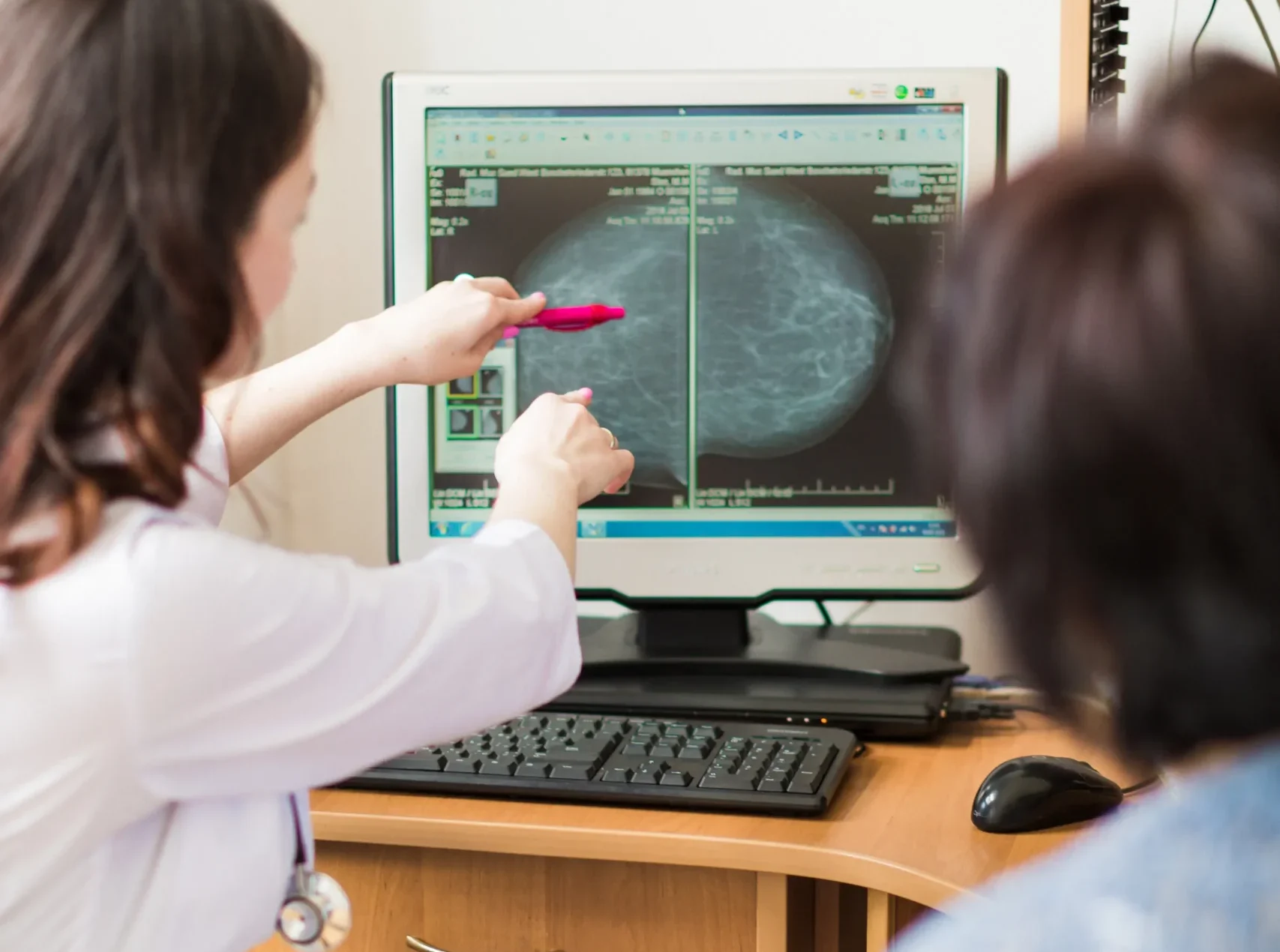
After your exam, your images are reviewed by Board Certified, Fellowship Trained radiologists with expertise in Breast Imaging. A report is then sent to the provider who ordered your ultrasound.
Your provider will review the results with you and explain any next steps. In many cases, that may simply mean returning to routine breast screening. If additional breast Imaging is recommended, your provider will explain what is being evaluated and why.
Frequently Asked Questions
Can you confirm breast cancer with an ultrasound?
Breast ultrasound can provide important information about a finding, but on its own it usually cannot “confirm” breast cancer. Ultrasound is very good at showing the characteristics of an area, for example, whether it looks fluid-filled (like a cyst) or solid, and whether it has features that may need further evaluation.
If something looks suspicious, the next step is often additional Breast Imaging or a biopsy, because a tissue sample is what confirms a diagnosis. Ultrasound is often part of that process, either to better understand a finding or to guide a biopsy when a sample is needed.
Should I worry if I need an ultrasound after a mammogram?
It is completely understandable to feel anxious, but needing an ultrasound after a mammogram is common and does not automatically mean something is wrong. Mammograms can show an area that needs a little more evaluation, and ultrasound is one of the best tools for answering certain questions, especially when the goal is to focus on a specific spot.
Many follow-up ultrasounds end up showing something benign, such as normal tissue overlap or a simple cyst. Your provider and the radiologist use the ultrasound findings to decide whether anything else is needed or whether you can return to routine screening.
Which is better, mammogram or breast ultrasound?
Neither one is “better” across the board because they are designed for different purposes. Mammography is the primary tool used for routine breast cancer screening and can detect certain changes, like calcifications, that ultrasound may not show as well.
Breast ultrasound is often used to evaluate a specific area or symptom and can help characterize certain findings in real time. In many situations, the most helpful approach is using them together, depending on what your provider is evaluating and what has already been seen on Breast Imaging.
Can I skip a mammogram and just do an ultrasound?
For most patients, ultrasound is not a substitute for a screening mammogram. Mammography remains the standard screening exam because it can detect some early changes that ultrasound may miss.
There are situations where ultrasound may be used as an additional exam, or where your provider may recommend a different approach based on your history, breast tissue, or specific symptoms. The best plan depends on your individual situation, so it is worth discussing with your provider rather than trying to choose one test on your own.
Does a breast ultrasound use radiation?
No. Breast ultrasound uses sound waves to create images and does not involve radiation.
Do I need an order from my provider?
In many cases, yes. Breast ultrasound is often scheduled based on an order from your provider, so the exam matches the reason it is being requested.
If you are unsure what was ordered or what you need to schedule, the Bay Radiology team can help you confirm the correct exam and guide you through the next step.
Questions About Your Breast Ultrasound?
If you have questions about an order, scheduling, or what to expect at your appointment, the Bay Radiology team is here to help.