Sometimes the next step in care is simply getting a clearer look. Ultrasound Imaging can do that without radiation, using sound waves to create real-time images that support diagnosis and follow-up care. Bay Radiology offers outpatient Ultrasound Imaging in Panama City, FL, for a wide range of common exams, from abdominal and thyroid studies to targeted follow-up when symptoms need answers.
Ultrasound images are interpreted by Board Certified, Fellowship Trained radiologists, and a report is sent to the provider who ordered your study to guide treatment.
Understanding Ultrasound Imaging
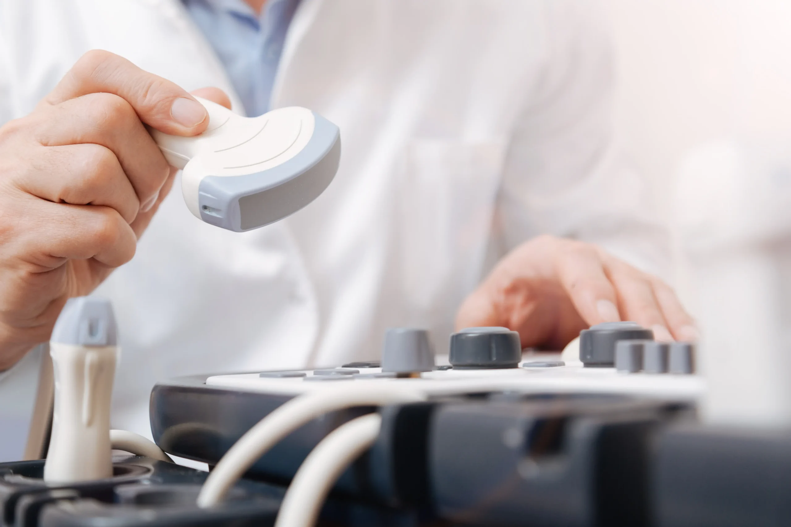
Ultrasound Imaging, also called sonography, works by sending high-frequency sound waves into the body using a small handheld probe placed on the skin. The sound waves bounce off internal structures and return to the probe, where a computer converts them into live images on a monitor.
This process allows providers to see organs, soft tissue, and blood flow during the exam itself. Since ultrasound does not use radiation and is typically very quick and comfortable, it is commonly used as an early step in evaluating many medical concerns.
Ultrasound Imaging Services
Bay Radiology offers several types of Ultrasound exams, from common diagnostic studies to more specialized Breast Imaging.

Standard Ultrasound
Used for common diagnostic studies such as abdominal, renal, pelvic, and thyroid exams.
Doppler Ultrasound
Adds blood-flow evaluation when circulation is part of what your provider needs to assess.
Breast Ultrasound
Often used to evaluate a specific area of concern after screening or a physical exam.
ABUS
Automated Breast Ultrasound that adds Imaging beyond mammography for dense breast tissue.
What to Expect During Your Ultrasound
Ultrasound exams are typically comfortable and straightforward. After check-in, a member of our Imaging team will guide you to an exam room and ask you to lie on a padded table. Depending on the type of study your provider ordered, you may be asked to lie on your back, side, or occasionally change positions during the exam.
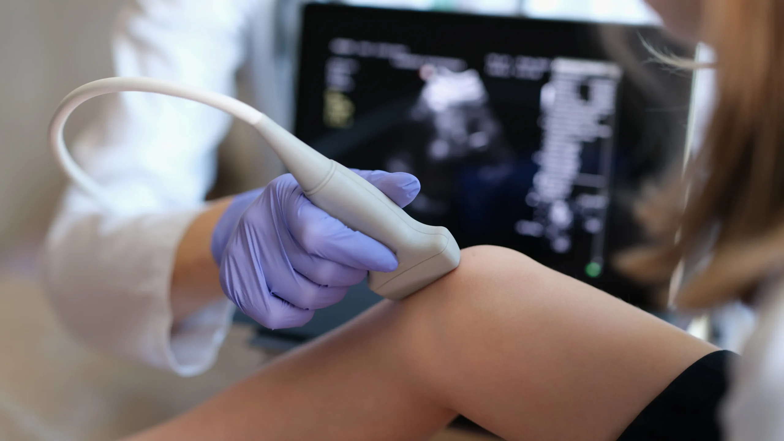
A small handheld probe is placed against the skin with a layer of gel that helps the device move smoothly and capture clear images. The gel is often warmed for comfort and wipes away easily after the exam.
As images are taken, you may be asked to hold your breath briefly or remain still for a moment while the images are captured. Most ultrasound exams are completed in a short visit, and you can return to normal activities right afterward.
How to Prepare
Preparation for an ultrasound is usually simple. Many exams require little or even no special preparation at all before your visit.
Here are a few general tips from our radiologists:
- Wear comfortable clothing that is loose-fitting around the area to be examined, or that can be rolled up.
- In many cases, you may not need to change clothes if the area is easily accessible
- Some ultrasound exams may require specific preparation, such as drinking water beforehand or avoiding food for several hours
- If special preparation is needed, your provider or our scheduling team will let you know in advance
View Preparation Guidelines
Results and Next Steps
Because Ultrasound is often used to take a closer look at a specific concern, the next step can vary depending on what the exam shows. After a radiologist reviews your images, a report is sent to your provider so they can discuss the findings with you and recommend any follow-up care or additional testing.
Frequently Asked Questions
How long does an ultrasound take?
Most ultrasound exams take about 20 to 30 minutes, though some may be shorter or longer depending on the area being examined. If your provider ordered multiple views or a more detailed study, your appointment may take a little more time.
Can urgent care do an ultrasound?
Some urgent care clinics offer limited ultrasound exams, but many do not have the equipment or radiologist support needed for a full diagnostic study. When more detail is needed, patients are often referred to an Imaging center like Bay Radiology, where the exam can be performed and interpreted as part of a larger care plan.
How does ultrasound work?
Ultrasound works by using sound waves instead of radiation. A small handheld probe sends sound waves into the body, and a computer turns the echoes that bounce back into live images on a screen.
Can ultrasound detect cancer?
Ultrasound can help identify areas that need a closer look, which is why it is often used in diagnostic follow-up. It can show whether something looks solid or fluid-filled and help guide next steps, but it does not confirm cancer on its own.
Is ultrasound radiology?
Ultrasound does not use ionizing radiation, so it is different from X-ray or CT in that sense. But it is still considered part of radiology because it is a medical Imaging exam that is commonly performed in Imaging centers and interpreted by radiologists.
What is a renal ultrasound?
A renal ultrasound is an ultrasound exam of the kidneys and nearby structures. Providers often order it to evaluate things like kidney stones, swelling, blockage, or other changes that may help explain symptoms.
What diseases can be detected by ultrasound?
Ultrasound can help detect or evaluate many conditions, depending on the area being examined. It is commonly used to look for things like gallstones, kidney stones, cysts, thyroid nodules, fluid collections, and other changes that help explain symptoms or guide follow-up care.