A mammogram is not something most women look forward to, but it is one of the most important screenings you can get. Even as new technologies have expanded the field of breast Imaging, mammography continues to be the foundation of breast cancer detection. It can reveal even minute changes before symptoms arise, when treatment is often simpler, and outcomes can be better.
At Bay Radiology, mammography is part of a comprehensive approach that includes both routine screening and diagnostic evaluation. We offer multiple mammography technologies so each exam can be matched to the patient, and every study is interpreted by Board Certified, Fellowship Trained radiologists with specialized expertise in breast Imaging.
What is Mammography?
Mammography is a specialized type of X-ray Imaging used to look at breast tissue in detail. It can identify small changes long before they would be noticeable during a physical exam, which is what makes it such a powerful screening tool.
Modern mammograms are digital, which allows radiologists to review breast tissue with greater clarity and precision. That detail helps support everything from annual screening to more focused follow-up when a question needs to be answered.
Screening and Diagnostic Mammograms
Not every mammogram is the same. Some are performed as part of routine annual screening, while others are ordered to take a closer look at a symptom or finding.

Screening Mammography
This is the type of mammogram most women have as part of regular breast cancer screening. It is done when there are no symptoms and no known problems.
Diagnostic Mammography
A diagnostic mammogram is a more focused exam used when there is a lump, pain, nipple discharge, a callback after screening, or another breast concern. It may include additional images to help the radiologist evaluate one area more closely.
Mammography Technology at Bay Radiology
Mammography has come a long way, and today’s technology offers more detail than ever before. Bay Radiology provides several advanced mammography options so screening and diagnostic exams can be performed with the Imaging approach best suited to the question being asked.
Traditional Digital Mammography
Modern digital mammography remains the foundation of breast cancer screening. It produces high-resolution breast images that radiologists can review with greater clarity than older film-based systems.
3D Mammography (Breast Tomosynthesis)
3D mammography captures the breast in thin layers rather than a single flat image. This added detail can make it easier to see through overlapping tissue and evaluate subtle findings more clearly.
Contrast-Enhanced Mammography
Contrast-enhanced mammography uses contrast material to highlight areas of increased blood flow within the breast. It adds another level of detail in certain diagnostic situations when standard mammography alone may not tell the full story.
When to Start Mammogram Screening
Most major medical organizations recommend annual mammogram screening starting at age 40. Having a mammogram every year supports early detection and has been shown to save more lives from breast cancer.
For some women, screening may begin earlier. Family history, genetic factors, prior breast conditions, or other health considerations can all influence when screening starts. This is why conversations with your provider are so important. Guidelines offer a helpful starting point, but screening decisions are always made with the individual patient in mind.
Staying consistent with recommended screenings matters because breast changes can develop over time. When mammograms are performed regularly, radiologists can compare images from year to year, making it easier to recognize subtle changes early.
When Additional Breast Imaging Is Needed
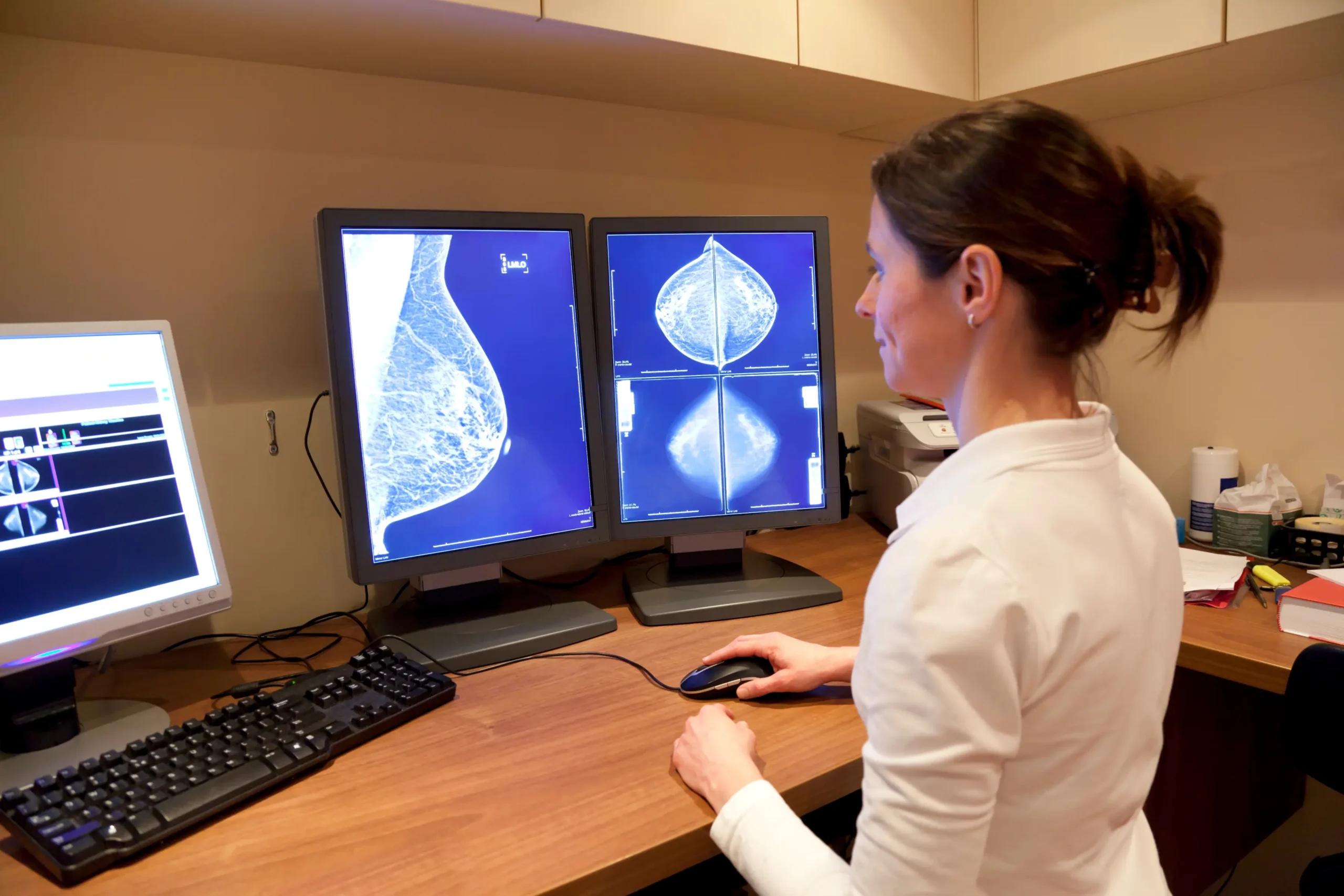
Mammography is the foundation of breast cancer screening, but it is not the only tool available for evaluating breast health. In some situations, your provider or radiologist may recommend another type of Breast Imaging to gather more information.
At Bay Radiology, several imaging options are available to support mammography and help provide a more complete picture when questions arise. These may include:
- Breast ultrasound, which can help evaluate certain breast findings
- ABUS (Automated Breast Ultrasound), often used as a supplemental screening option for patients with dense breast tissue
Having multiple Breast Imaging tools available helps ensure that changes in breast tissue can be evaluated thoroughly and as early as possible.
What to Expect During a Mammogram
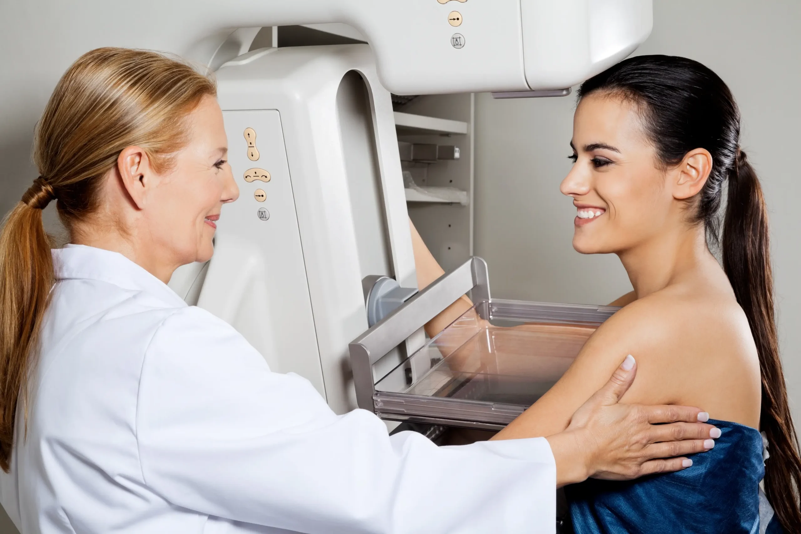
During a mammogram, you will stand in front of the imaging system while a member of the Breast Imaging team gently positions the breast on a flat imaging plate. The breast is briefly compressed between two plates so clear images can be captured.
Compression can feel uncomfortable for a few seconds, but most patients find it more tolerable than they expected. The technologist will guide you through each step, explain what is happening, and take the images as quickly as possible while still ensuring accuracy.
Images are usually taken from a few different angles, and the entire exam typically takes about 15 to 20 minutes. Advances in digital mammography technology have helped improve image clarity while also keeping exam times short.
Your privacy and comfort are always respected during the exam, and the team works carefully to make the experience as smooth and supportive as possible.
How to Prepare
Preparing for a mammogram is simple, but a few small steps can help ensure clear images and a smooth visit.
- Avoid deodorant, lotion, powder, or perfume on the day of your exam, as these can appear on the images.
- Wear a two-piece outfit so it is easy to change for the exam.
- Let the technologist know if you have any breast symptoms or concerns.
- If you have had mammograms at another facility, bringing those prior images can help the radiologist compare results over time.
See More Preparation Tips
Results and Next Steps
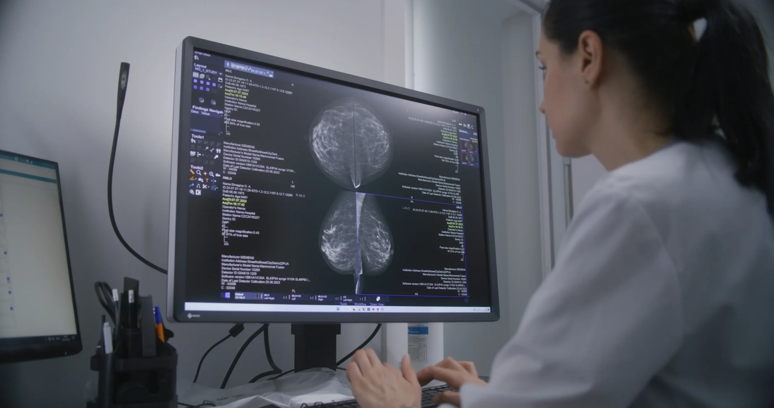
After your exam, the images are reviewed by radiologists with expertise in Breast Imaging. A report is then sent to the provider who ordered your study.
In many cases, the next step is simply continuing routine screening on the schedule recommended by your provider. If the radiologist sees something that should be followed more closely, your provider may recommend additional Breast Imaging or short-term follow-up. Your provider will discuss the results with you and explain what comes next.
Frequently Asked Questions
What is a diagnostic mammogram?
A diagnostic mammogram is a type of mammogram used when there is a specific concern to evaluate. This might include a breast symptom such as a lump or pain, or a follow-up after a screening mammogram that needs a second look. Diagnostic mammograms often include additional images focused on a particular area so the radiologist can review the tissue more carefully.
How long does a mammogram take?
The imaging portion of a mammogram usually takes only a few minutes, and most appointments are finished within about 15 to 20 minutes. Your technologist will guide you through the process step by step to make sure the images are clear while keeping the exam as efficient and comfortable as possible.
What does breast cancer look like on a mammogram?
Breast cancer can appear in different ways on a mammogram. Radiologists look for patterns such as small white spots called calcifications, areas of dense tissue, or masses that have irregular shapes. Not every unusual finding is cancer, which is why mammograms are interpreted by experienced radiologists who carefully review the images and determine whether additional Imaging is needed.
What age should you get a mammogram?
For most women at average risk, annual mammogram screening begins at age 40. If you have higher-risk factors, such as a strong family history or certain genetic risks, your provider may recommend starting earlier or adding other Breast Imaging as part of your screening plan.
Does a mammogram hurt?
Mammograms can feel uncomfortable for a few seconds because the breast must be gently compressed to capture clear images. Most patients describe the sensation as pressure rather than pain, and it lasts only briefly while the image is taken. The technologist works carefully to position the breast and complete the exam as quickly as possible.
How does a mammogram work?
A mammogram uses a low-dose X-ray system designed specifically for breast tissue. During the exam, the breast is positioned on an imaging plate and briefly compressed so detailed images can be captured from different angles. These images allow radiologists to examine breast tissue closely and identify changes that may need further attention.
What is the difference between mammography and tomosynthesis?
Mammography is the general term for breast X-ray imaging. Tomosynthesis, often called 3D mammography, is an advanced form of mammography that captures multiple images of the breast from different angles. These images are combined to create thin “slices” of breast tissue, which can make certain findings easier to see.
Can you get a mammogram while pregnant?
Yes. Mammography is safe during pregnancy and breastfeeding, and routine screening should continue for both average-risk and high-risk women when it is age-appropriate. Diagnostic mammography and/or ultrasound may also be used to evaluate breast symptoms during pregnancy. Always let your care team know if you are pregnant or think you might be before any Imaging exam.
Reference: ACR Appropriateness Criteria® Breast Imaging During Pregnancy (JACR)
Does a painful mammogram mean cancer?
No. Discomfort during a mammogram is related to the brief compression used to capture clear images and does not indicate cancer. Many women experience some pressure during the exam, especially if the breasts are sensitive at that time of the month. If you have concerns about breast pain outside of the exam, it is always a good idea to discuss them with your provider.
Questions About Mammography or Breast Screening?
If you have questions about screening guidelines, dense breast tissue, or the type of exam your provider recommended, our team is here to help you understand your options.