July 17, 2026
Breast Ultrasound vs Mammogram: Which is Better?
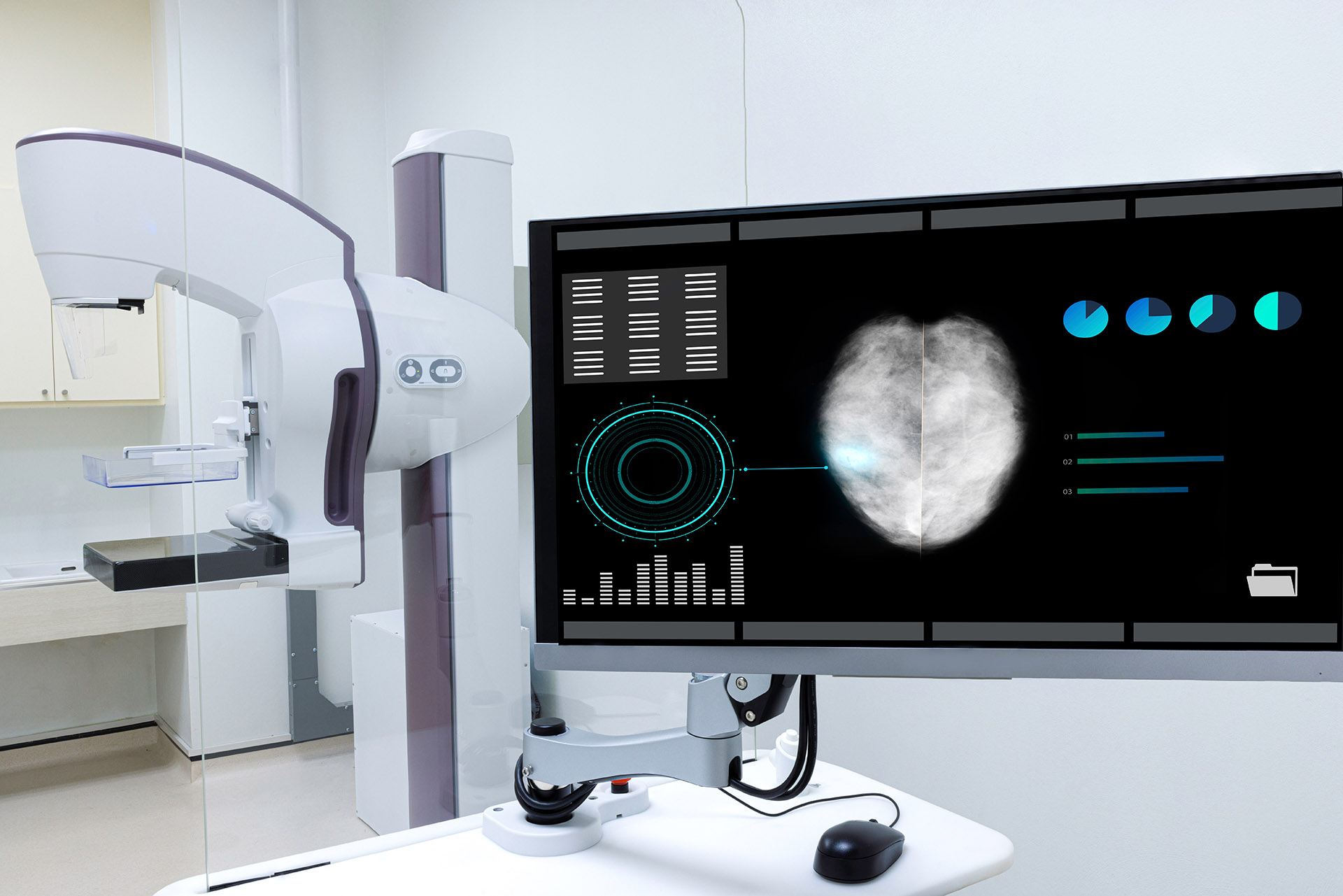
A breast ultrasound can sound like it would be a much easier annual exam than a mammogram. There is no breast compression, no radiation, and for many people, the whole idea feels less intimidating. So when a doctor recommends a mammogram, it makes sense to wonder: why can’t an ultrasound do the job instead?
Because these tests aren’t interchangeable. They’re built to detect different kinds of breast changes, and sometimes the most responsible answer is using both.
Can a Breast Ultrasound Replace a Mammogram?
For most patients, no. A breast ultrasound should not be used instead of a mammogram for routine screening.
The reason is that mammography and breast ultrasound have different purposes in Breast Imaging.
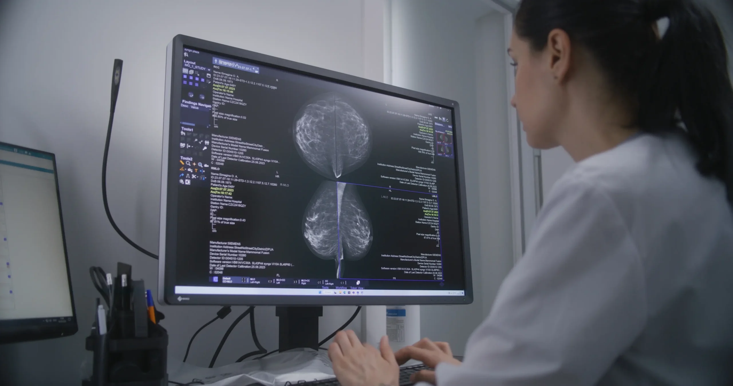
Mammography is typically used as a screening tool. It looks across the breast for subtle changes that may be too small to feel and may not be seen clearly with ultrasound.
Breast ultrasound is typically used as diagnostic or supplemental Imaging. A radiologist may use it to take a closer look at a specific area, such as a finding seen on a mammogram or a lump that needs further evaluation.
Ultimately, breast ultrasound can be very helpful when used alongside mammography. But it does not replace regular screening mammograms.
Breast Ultrasound vs Mammogram: A Simple Comparison
A mammogram and a breast ultrasound use different technology to create images, which is why they’re used in different situations.
- A mammogram uses low-dose X-rays. The breast is briefly compressed so the tissue can be spread out and seen more clearly.
- A breast ultrasound uses sound waves. A handheld device is used to scan the area being evaluated without compression.
Here is a simple side-by-side comparison:
| Question | Mammogram | Breast Ultrasound |
|---|---|---|
| How does it work? | Uses low-dose X-rays | Uses sound waves |
| Uses breast compression? | Yes | No |
| Common role | Routine breast cancer screening | Diagnostic or supplemental Imaging |
| Looks at | The breast broadly for early changes | A specific area or concern in more detail |
Does Mammogram Radiation Make Ultrasound “Safer”?
This is a common worry, and it makes sense to ask.
Ultrasound does not use radiation. Mammography does use low-dose X-rays, but the dose is small and tightly regulated. Mammograms are widely recommended because the benefit is early detection, finding changes when they’re smaller, treatment can be simpler, and outcomes are often better.
If mammography were replaced with ultrasound alone, some early findings that mammograms are designed to detect could be missed until they become larger or easier to feel. So the question isn’t only “which test has less radiation,” it’s which test is most likely to catch important changes early, when it matters most.
What a Mammogram Can Show That Ultrasound May Miss
Mammograms are especially important because they can show subtle changes in breast tissue before a lump can even be felt.
One key example is calcifications. Calcifications are tiny calcium deposits in the breast. Many are benign, but certain patterns can be an early sign of breast cancer or precancerous change. Because calcifications are usually seen on mammography, a mammogram can help identify concerns that ultrasound may not show.
Breast compression also serves an important purpose. While it can be uncomfortable, it helps spread out the tissue so the radiologist has a clearer view. When tissue overlaps, small findings can be harder to see or may be hidden by other tissue. This is especially true when dense breast tissue is present.
For some patients, 3D mammography may provide even more detail. It creates layered images of the breast, which can help the radiologist review areas of tissue that may overlap on a standard mammogram.
What Breast Ultrasound Is Best For
Breast ultrasound has a different strength than mammography. Instead of showing subtle changes, ultrasound can help evaluate something that has already been seen or felt. That may include an area noted on a mammogram, a lump found during a breast exam, or even an area where tenderness is present.
One of the most helpful things ultrasound can do is show whether something looks:
- Fluid-filled (often a cyst, which is common and usually benign), or
- Solid (made of tissue, which may still be benign but often needs a clearer explanation)
This distinction matters because a lump can feel the same from the outside whether it is fluid-filled or solid. Ultrasound helps give the radiologist more information about what is happening inside the breast and what the next step should be.
Why Do People Get an Ultrasound After a Mammogram?
Getting a breast ultrasound after a mammogram is very common, and it doesn’t automatically mean cancer. It also doesn’t mean the mammogram “didn’t work.”
In many cases, the mammogram did exactly what it’s supposed to do: it picked up a small change or an area that deserves a second look with a different type of imaging. Ultrasound helps answer the next question: what that area most likely represents.
Sometimes the follow-up ultrasound shows something simple, like a cyst. Sometimes it helps confirm that a spot is just normal tissue. And sometimes it helps determine whether additional imaging or a biopsy is the right next step.
What If You Have Dense Breasts?
Dense breast tissue is common, and it changes how a mammogram looks. On a mammogram, dense tissue appears white, and many concerning findings can also appear white. That “white on white” overlap can make it harder to spot small changes in dense breasts.
This does not mean mammograms aren’t useful. It means your doctor may recommend additional Breast Imaging based on your results and personal risk factors.
Related Reading: What Dense Breast Tissue Means
How to Know What’s Right for You
Mammograms are still the foundation of breast cancer screening, but they aren’t the only tool your care team may use. Ultrasound is one of several imaging options that can be added when your history, breast density, or symptoms call for it. If you have questions about why a test was recommended, bring them to your doctor. The “right” plan is the one that fits you.
Related Articles